
Sport injuires:
The lesion of the anterior cruciate ligament
General information of the physiotherapy after LCA reconstruction in the hospital:
Please click here
Model Physiotherapy 1:
Please click here
Model Physiotherapy 2:
Please click here
Index
1. Introduction
2. Rupture of the Anterior Cruciate Ligament
2.1 Function and Anatomy of the Anterior Cruciate Ligament
2.2 Mechanism of Injury
2.3 Diagnosis
2.4 Treatments
2.4.1 Knee Arthroscopy
2.4.2 Treatment of the Rupture of the Anterior Cruciate Ligament
2.4.2.1 Conservative Treatment
2.4.2.2 Operative Treatment
2.4.2.3 Post-Operative Video Recording
2.4.2.4 Information on Physiotherapy for ACL Patients
2.4.2.4.1 Post-Treatment Plan for In-Patient Physical Therapy After ACL Surgery (with Semitendinous-Gracilis Double Loop Technique)
2.4.2.4.2 Post-Treatment Plan for Out-Patient Physical Therapy After ACL Surgery (with Semitendinous-Gracilis Double Loop Technique)
1. Introduction
With the increasing number of participants in athletics, the number of sports injuries is steadily climbing. After traffic accidents, sports injuries are the most frequent causes of ligament injuries to the knee joint. In sports injuries, the knee is generally the most commonly affected joint. The bone, whose basic framework breaks only under very strong forces, is much less frequently affected in typical sports injuries. The soft parts of the knee joint are much more frequently affected by accidental injuries or overloading injuries. The soft parts include the collateral and cruciate ligaments, which are responsible for stability; the menisci, which control knee alignment; and the tendons and their bursa, which create the active course of movement.
The knee cartilage, which coates the bones of the joint as a sliding layer and which is responsible for the friction-free movement and springiness of the joint, is another structure that can be damaged either acutely or chronically. Knee joints are especially prone to injury. This is based in the fact that, first of all, the human being from an evolutionary perspective has "only recently" stood on two legs. Another cause for the increasing frequency of knee injuries is based in the fact that in recent years amateur sports have rapidly increased. In addition to the classic contact sports like football, handball, and ice hockey, the knee is also at risk in some types of non-contact sports, such as skiing, tennis, and even dancing. Ever more extreme and accident-susceptible types of sports have been added to the classical sports (e.g., snow boarding, in-line skating, mountain biking); they are enjoying ever greater popularity. In the traditional sports, increased training and equipment demands lead to an increased risk of accidents (e.g., ambitious amateur football players must cover running distances in a competitive match that a few years ago would have been managed only by football professionals). For example, improved skiing equipment has resulted in a faster descent and consequently to an increased risk of injury. In the meantime, ski boots protect the ankle joint and have shifted accidents to the knee joint. This improved equipment and increased speed bring about greater forces on the musculo-skeletal system that is not always tolerated and can lead to severe injuries, such as a rupture of the anterior cruciate ligament.
2. Rupture of the Anterior Cruciate Ligament
A tear of the anterior cruciate ligament is a severe injury. Until a few years ago, such an injury in sports meant the end to an athlete's career. Thanks to ever increasing knowledge of the anatomy, the physiopathology, and the biomechanics of the knee joint, thanks to a more refined set of instruments and to modern operative techniques, and thanks to differentiated follow-up treatment, patients today can again be made fit for sports.
2.1 Function and Anatomy of the Anterior Cruciate Ligament
The knee joint must fulfill two tasks that are scarcely compatible. On the one hand, it must assure maximum stability, while on the other it must give maximum mobility. For these tasks the human knee has several structures – for example, the musculature (active stabilization) does the stabilizing, guiding the knee joint dynamically and actively both in everyday activities as well as in sports, and the "passive structures," guaranteeing stability under dynamic and static conditions. The passive structures include the menisci, the lateral ligaments, the capsule, the cartilage cover of the joint surfaces, and, as the central support pillar of the knee joint, the anterior and posterior cruciate ligaments.
The anterior cruciate ligament (ACL) is formed by two bundles of ligaments, the anterior-medial and the posterior-lateral bundles. The ACL in the main consists of firm connective tissue (type I collagen, type III collagen). It lies in the center of the knee joint and forms a connection between the upper and lower leg bones (see Illustration 1).

(Illustration 1): View of a bent left knee joint from the front with presentation of the ligaments and the distal part of the upper leg. The patella (kneecap), the musculature, and the joint capsule have been removed for a better view. The fiber bundles of the cruciate ligaments are clearly visible.
The main function of the ACL is to secure the lower leg in relation to the upper leg against a forward displacement. This task is done most effectively at a bending of the knee joint of 20°-30°. The cruciate ligament does not have only mechanical stabilization functions, as previously assumed. As known today, the cruciate ligament has sensory functions as well as mechanical functions. Nerve receptors are located in the areas of the ligament-bone intersection (insertion area) and in the skin stretched over the ligament (synovia); these receptors deliver information to the nervous system regarding the position of the knee joint and the tension of the cruciate ligament.
This information is forwarded further to the muscles of the knee joint. Studies have shown that after a rupture in the cruciate ligament, the innervation of the musculature of the affected extremity, particularly of the knee joint extensors, is disturbed. This is evident in a weakening of the muscles and in problems of coordination, that is, patients with a tear in the anterior cruciate ligament have clear problems in perceiving the position of the knee joint.
2.2 Mechanism of Injury
The typical pattern of injury originates in a sudden strong outward twisting of the lower leg at the same time as a bending of the knee joint. (A typical example is the tip of the foot getting caught in full stride, or while threading the tip of the ski through a slalom gate). In these cases the knees are in bent position, and the lower leg is suddenly turned strongly to the outside by a lever arm, such as a ski.
In the newer sports like snowboarding, in-line-skating, and mountain biking, other injury mechanisms are known as well. In these there are various mechanisms that lead to a rupture in the anterior cruciate ligament. In general, ligament injuries occur when our body's protective reflexes set in too late. That means that the active stabilization of the knee joint through its muscles requires a certain reaction time (approx. 0.13 seconds) in order to compensate for the stretching of the muscles by the forces affecting the knee. In sport accidents, there is a very "rapid" high force exerted (0.05 seconds), and as a result the defense mechanism set in motion by the tendon and muscle reflexes comes too late, and the entire force of the external pressure comes to bear directly on the ligaments, which do not withstand this force. Frequently, however, other structures of the knee joint are affected as well.
In particular the lateral ligaments, the menisci, the structures of the joint capsule, and the cartilage are also affected. An important companion injury to a rupture of the anterior cruciate ligament is the injury to the posterior and lateral knee joint structures (so-called posterior-lateral instability). This is an important injury, since it is often not diagnosed, but it can be responsible for a residual instability of the knee joint after a successful operation. Ligament tears do not obey the "all-or-nothing principle." Depending on the force exerted, the damage can be light or severe.
2.3 Diagnosis
Usually the joint fills with blood after a fresh injury, and results in a marked swelling of the knee joint. The swelling results in the inability to fully bend/extend the knee joint. In most cases, the knee joint is painful, but occasionally ruptures of the cruciate ligament occur without perceptible pain, especially if they occur during a sport match, in which the athlete is under stress and distracted. Most patients complain of a subjective feeling of instability in the affected knee joint ("giving way"). They have a feeling of insecurity, since they may no longer be able to stabilize the knee joint, due to the failing functioning of the cruciate ligament; in certain movements it bends away uncontrollably (what is called the "pivot shift" phenomenon).
The diagnosis of anterior cruciate ligament rupture is in most cases made clinically. In addition to close questioning of the patient about the cause of the accident and an assessment of the symptom profile (swelling, pains, feeling of instability), a precise examination with specific examination techniques leads to the diagnosis by the physician. A reliable diagnosis can clinically be made with an increased forward movability of the lower leg in relation to the upper leg as compared to the other side, with a gentle tap of the cruciate ligament (so-called positive Lachmann test), and with a positive Macintosh pivot shift test (the so-called rotation-point-slide sign).
Various procedures are available today to be sure of the diagnosis. The conventional X-ray image is a diagnostic standard, probably more to exclude knee joint fractures than to show a rupture of the cruciate ligament. Needle tapping of a joint is indicated for strong swelling of the traumatized knee in order to remove the fluid from the knee joint and thus to relieve the knee joint of stress. Needle tapping secondarily gives information about the type of injury; if the physician draws pure blood from the knee joint, then an injury to the cruciate ligament is quite likely. However, if globules of fat are found along with blood during the tapping, then one must assume a bone injury.
2.4 Treatments
2.4.1 Knee Arthroscopy
Diagnostic knee arthroscopy (knee imaging) is an operation that is done both under partial and full anesthesia. The patient can follow the entire operation on video and also be informed of the intra-operative findings and of the reconstruction involved in the cruciate ligament surgery (Illustration 2).
In this, a camera and two trocars are inserted into the joint through three small skin incisions (this is the way the instruments needed for the operation are inserted into the knee joint). Arthroscopy has the advantage of allowing the entire knee joint to be observed from the inside by the operator. Any fluids can be drained and injuries immediately dealt with. This applies in particular to additional injuries, such as a meniscus lesion, for which the quickest possible intervention is of great importance, as well as to the rupture of the cruciate ligament.

(Illustration 2): Diagnostic knee arthroscopy (knee imaging): On the left the bent knee joint; on the right, the television image for the operator and for the patient.
2.4.2 Treatment of the Rupture of the Anterior Cruciate Ligament
Basically, there is a choice of treatment for injury to the anterior cruciate ligament between conservative and operative therapy. Which approach is taken depends primarily on the athletic ambitions of the patient, and secondarily on the patient's age and the demands made by everyday life and occupation. Clinical results of conservative therapies in most cases give functionally unsatisfactory results, and the patients complain of persistent feelings of instability and of knee pains. Most of these patients very quickly develop degenerative changes and additional knee joint injuries, such as meniscus tears or other ligament lesions.
2.4.2.1 Conservative Treatment
A knee joint without an anterior ligament is usually adaptable to the demands of everyday living. Conservative therapy comes into play for patients who have no interest in competitive sports or in general engage in little sports activity, can adjust and reduce their activity levels, or are over 60-65 years old. The biological age of the patient plays an important role. Physiotherapy helps to compensate for the missing cruciate ligament through planned build-up of the muscles and through coordination training. What is important here is a highly motivated patient and insight into the necessity of regular exercise. Problems appear as soon as the muscles can no longer compensate for forces appearing in sports or in everyday or occupational activities, and the patient thus detects a feeling of instability in the knee joint ("giving way" of the knee joint). If this is the case, the conservative therapy should be reevaluated, and there should be no delay in stabilizing the knee joint operatively with a substitute cruciate ligament. Today we know that a chronically unstable knee joint can lead to addition damage to the meniscus and cartilage, which promotes the premature wearing away of the joint.
2.4.2.2 Operative Treatment
The tear of the anterior cruciate ligament indicates a quickly scheduled arthroscopically assisted surgery with a cruciate ligament substitute if the patient desires to continue his sports ambitions. The modern fixation methods of a substitute cruciate ligament allow quick weight bearing when accompanied by physiotherapy, mobilization, and load bearing immediately following the operation . In addition to what is called open cruciate ligament substitute surgery (in which the new cruciate ligament can be inserted by a mini-arthrotomy in the open knee joint) the arthroscopically assisted substitute anterior cruciate ligament surgery has today become established as the standard for the operative treatment of an ACL tear. Thanks to the great potential for a reintegration of the cruciate ligament transplant into the joint and because of the failure of the prosthetic substitute ligament, today biological tissue transplants are used for cruciate ligament surgery. Biological tissue transplants are available in the form of autograft (from the individual himself) and allograft (from a person of the same type). For what are called autograft tissue transplants, the middle third patellar tendon, the doubled semitendinous and gracilis tendons, and the quadriceps tendons can be used. We do not use allografts because of the risk of infection. The quadriceps tendon is used primarily for repair of a repeat anterior ligament injury and in multi-ligament injuries. Essentially, two methods are available to choose from, depending on the choice of the transplant. The middle third patellar tendon with two bone blocks (Illustration 3) or the semitendinous-gracilis tendon is taken from the patient (see Illustration 4).

(Illustration 3): The middle third patellar tendon (center) with two bone blocks (on either end) is shown.

(Illustration 4): The removal site is shown with a short incision of the semitendinous and gracilis tendon.
The middle third patellar tendon transplant has become very popular, since the transplant has shown a highly stretchable load ("ultimate tensile load") of 2376 N (as opposed to an intact ACL with 2160 N) and a high degree of stiffness of 620 N/mm (versus an intact ACL of 242 N/mm) and since the transplant, with the help of two bone blocks, can be fixed very tightly to the bone. Because of knee pains, weakness in the extensor apparatus, decreased knee mobility, and risk of fracture of the patella, the doubled semitendinous-gracilis tendon transplant has grown in importance in recent years. The advantages of this transplant are above all the absence of knee pains after the operation and a rapid full-range movement of the knee joint. In addition, this transplant shows a very high stretchable load of 4108 N (as opposed to an intact ACL with 2160 N).
The doubled semitendinous-gracilis tendon transplant also exhibits stiffness similar to that of a healthy ACL, while the middle third patellar tendon is 3 to 4 times stiffer. Primarily in the middle third patellar tendon, a too strong tension of the transplant results in reduced mobility of the knee joint, and regaining the maximum mobility of the knee joint takes longer. The disadvantages of the doubled semitendinous-gracilis tendon transplant were until recently an inadequate fixation of the transplant in the bone and delayed healing in the bony canals.
Today, due to the improvement of the fixation method, e.g., by Intrafix fixation, and thanks to the off-center fixation close to the joint of the four tendon bundles in the bony canal, substitute cruciate ligament surgery with this transplant is very reliable and comparable to that of the middle third patellar tendon. There are even more recent studies that show that the maximum load up to failure of a doubled semitendinous-gracilis tendon transplant is much greater than that of a middle third patellar tendon (2422 N versus 1784 N).
With arthroscopy, the knee joint is prepared for the insertion of the cruciate ligament. For this, the torn cruciate ligament is removed and a canal for the "new" ligament is bored in the lower leg and the upper leg bones. Today, arthroscopy allows a visual, precise placement of the transplant, so that it comes to lie in the anatomic course (i.e., in the position of the old cruciate ligament).
The two methods are distinguished principally with regard to the fixation of the transplant. In the method that uses the patellar tendon as a transplant, the fixation of the femoral and tibial bone blocks is carried out with a titanium interference screw for each. This screw is implanted for life. However, this method is contra-indicated in patients with known pre-existing femoropatellar pains (knee pains in the patellar area, common in patients whose occupation involves a lot of kneeling, such as tile setters), for around 10% of the patients after the operation complain of persistent pains in the area where the bone block was removed, that is, the lower end of the patella.
For these patients, the second method described above is recommended, with the doubled semitendinous and gracilis tendon used as a ligament substitute, since here problems at the tendon removal site hardly ever occur. Wit the new fixation methods with RIGIDFIX® (fixation of the femoral portion, see Illustrations 5 and 6), and with INTRAFIX® (fixation of the tibial portion, see Illustrations 7-9), today one can achieve a very good primary fixation of the doubled semitendinous and gracilis tendon, and the patient can be mobilized and bear weights very quickly, as with the previously discussed method with the middle third patellar tendon. It is important to observe that today the results after an anterior cruciate ligament surgery with doubled semitendinous and gracilis tendon and with the middle third patellar tendon are equally good. Both methods allow the patient to resume competitive sports with a well rehabilitated muscle apparatus after 6 months.


(Illustration 5, live) and (Illustration 6, schematic): The new fixation methods with RIGIDFIX® (fixation of the femoral part): The RigidFIX system allows a very strong fixation of the transplant circumferentially in the bored bony canal in the femur with two bio-resorbable PLA cross pins of 3.3mm x 42mm diameter/length.

(Illustration 7, live): The new fixation method with BIO-INTRAFIX® of the doubled semitendinous and gracilis tendons (fixation of the tibial part) allows very rapid rehabilitation with increasingly full weight bearing.

(Illustration 8, schematic): The new fixation method with BIO-INTRAFIX® allows an off-center fixation of the individual semitendinous and gracilis tendons (fixation of the tibial part) in the bony canal.

(Illustration 9, schematic): The BIO-INTRAFIX consists of two resorbable components (TCP and PLA): a screw and a sheath with BIOCRYL material with an osteoconductive function. This fixation method allows a fixation strength of 1067 N as opposed to 800 N for an interference screw in the patellar tendon transplant.
(Illustration 10, arthroscopic finding):Fresh rupture of the anterior cruciate ligament in the center third (left in the picture, **) and an intact posterior cruciate ligament (right in the picture, *).
(Illustration 11, arthroscopic finding): Anterior cruciate ligament transplant with doubled semitendinosus and gracilis tendon (***).
2.4.2.3 Post-Operative Video Recording
In the following video recordings, there is the possibility of seeing some examples following arthroscopically assisted ACL surgery. (Here I would like to again thank the individual patients for their kind permission.)
- Patient A: 0 days after arthroscopically assisted ACL surgery on the right with the doubled semitendinous and gracilis tendon technique: Proximal fixation to the femur with RIGIDFIX® fixation method and BIO-INTRAFIX® distal fixation method to the tibia.
Please click here
- The same patient A: 2 days after arthroscopically assisted ACL surgery on the right with the doubled semitendinous and gracilis tendon technique: Proximal fixation to the femur with RIGIDFIX® fixation method and BIO-INTRAFIX® distal fixation method to the tibia.
Please click here
- The same patient A: 4 days after arthroscopically assisted ACL surgery on the right with the doubled semitendinous and gracilis tendon technique: Proximal fixation to the femur with RIGIDFIX® fixation method and BIO-INTRAFIX® distal fixation method to the tibia.
Please click here
- Patient B: 4 days after arthroscopically assisted ACL surgery on the right with the doubled semitendinous and gracilis tendon technique: Proximal fixation to the femur with RIGIDFIX® fixation method and BIO-INTRAFIX® distal fixation method to the tibia.
Please click here
- Patient C: 4 days after arthroscopically assisted ACL surgery on the right with the doubled semitendinous and gracilis tendon technique: Proximal fixation to the femur with RIGIDFIX® fixation method and BIO-INTRAFIX® distal fixation method to the tibia.
Please click here
- The same patient C: 2 weeks after arthroscopically assisted ACL surgery on the right with the doubled semitendinous and gracilis tendon technique: Proximal fixation to the femur with RIGIDFIX® fixation method and BIO-INTRAFIX® distal fixation method to the tibia.
Please click here
- Patient D: 1 year after arthroscopically assisted ACL surgery on the left with the doubled semitendinous and gracilis tendon technique: Proximal fixation to the femur with RIGIDFIX® fixation method and BIO-INTRAFIX® distal fixation method to the tibia. The patient can carry out his sports activities without limitation.
Please click here
- Patient E: 2 days after arthroscopically assisted ACL surgery on the right with the doubled semitendinous and gracilis tendon technique: Proximal fixation to the femur with RIGIDFIX® fixation method and BIO-INTRAFIX® distal fixation method to the tibia.
Please click here
- Patient F: 6 weeks after arthroscopically assisted ACL surgery on the left with the doubled semitendinous and gracilis tendon technique: Proximal fixation to the femur with RIGIDFIX® fixation method and BIO-INTRAFIX® distal fixation method to the tibia.
Please click here
- Patient G: 1 day after arthroscopically assisted ACL surgery on the left with the doubled semitendinous and gracilis tendon technique: Proximal fixation to the femur with RIGIDFIX® fixation method and BIO-INTRAFIX® distal fixation method to the tibia.
Please click here
- The same patient G: 2 days after arthroscopically assisted ACL surgery on the left with the doubled semitendinous and gracilis tendon technique: Proximal fixation to the femur with RIGIDFIX® fixation method and BIO-INTRAFIX® distal fixation method to the tibia.
Please click here
- Patient H: 2 days after arthroscopically assisted ACL surgery on the left with the doubled semitendinous and gracilis tendon technique: Proximal fixation to the femur with RIGIDFIX® fixation method and BIO-INTRAFIX® distal fixation method to the tibia.
Please click here
- Patient I: 2 days after arthroscopically assisted ACL surgery on the left with the doubled semitendinous and gracilis tendon technique: Proximal fixation to the femur with RIGIDFIX® fixation method and BIO-INTRAFIX® distal fixation method to the tibia.
Please click here
2.4.2.4 Information on Physiotherapy for ACL Patients
Here you can downlow information's of the physiotherapy after LCA reconstruction as pdf document.
General information of the physiotherapy after LCA reconstruction in the hospital:
Please click here
Model Physiotherapy 1:
Please click here
Model Physiotherapy 2:
Please click here
Beim Lesen dieser Informationen werden Sie erfahren, was Sie in den nächsten Tagen von unserer Seite her erwartet, welches die Schwerpunkte der Nachbehandlung sind und wie Sie selber Einfluss auf einen möglichst optimalen Wund- heilungsverlauf nehmen können.
Nur durch eine gemeinsame Anstrengung können wir ein optimales Resultat erreichen.
Nun wünschen wir Ihnen eine erfolgreiche Operation und Freuen uns auf eine gute Zusammenarbeit.
Der Eintrittstag
Sie werden gegen Abend von einer TherapeutIn unseres Physiotherapie-Teams be- sucht. Dabei werden Sie kurz über die Vorgeschichte des Knies befragt. Ihr Knie wird auf Beweglichkeit getestet, es werden für Sie die Gehstöcke angepasst und Sie er- halten erste Instruktionen, wie man mit den Gehstöcken geht und sich damit auf der Treppe zurecht findet.
Weiter werden Ihnen Atemübungen gezeigt, die nach der Operation zu einer be-
wussten Atemvertiefung führen sollen, um mögliche Komplikationen (z.B. Lungen-entzündung) zu vermeiden.
Der Operationstag
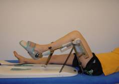
Wir werden Ihnen eine Bewegungsschiene (Kinetek) zur Erhaltung der Beweglichkeit anpassen. Auf dieser Bewegungsschiene wird ihr Knie passiv (d.h. ihre Muskulatur sollte dabei locker bleiben) dreimal täglich für eine Stunde gebeugt und gestreckt. Bitte beginnen sie langsam den Bewegungswinkel zu erhöhen, sodass sie keine Schmerzen, jedoch ein erhöhtes Spannungsgefühl wahrnehmen, welches Sie ertragen können.
Zusätzlich werden wir Ihnen Atemübungen zeigen, welche Sie stündlich 5x wiederholen (selbständig) sollten.
Der 1.Tag nach der Operation
Grossen Wert nach frisch operierten Kniegelenken wird auf die aktive Kniestreckung gelegt. D.h. es ist wichtig, dass Sie das Anspannen des vorderen Oberschenkelmuskels (M. Quadriceps) erlernen und auch trainieren werden. Die PhysiotherapeutIn wird Ihnen dazu verschiedene Übungen instruieren und Sie bitten, die täglich mehrmals zu machen. Dadurch erreichen Sie ein stabiles und sicheres “Knie-Gefühl“ beim Gehen.
Diese Kniestreckung, die auch beim Gehen eine grosse Bedeutung hat, werden wir (falls notwendig) immer wieder bei verschiedenen Gangübungen erwähnen und auch trainieren.
Am ersten Tag nach der Operation werden Sie sitzen können und wenn es die Kräfte erlauben, dürfen Sie das erste Mal aufstehen und die ersten Schritte an Gehstöcken oder an anderen Hilfsmitteln gehen (sofern das Gefühl der Beine vorhanden ist).
Das operierte Bein wird dazu bis über das Kniegelenk eingebunden, das „gesunde“ Bein bis auf Kniehöhe. Dies dient der Kreislaufunterstützung.
Bis zum fünften postoperativen Tag, sollten Sie schauen, dass das operierte Bein beim Gehen und beim Sitzen bis über das Kniegelenk einbandagiert ist, um eine Schwellung zu vermeiden.
Die Belastungslimite (in kg) des operierten Beines bestimmt der Operateur. Die zu-ständige PhysiotherapeutIn wird Sie diesbezüglich informieren und instruieren.
Der 2.Tag nach der Operation
Neben der aktiven Kniestreckung ist auch die Beugung sehr wichtig. Beugen Sie das operierte Bein mehrmals am Tag möglichst schmerzfrei. Dabei ist zu beachten, dass die Ferse immer Kontakt zur Bettunterlage behält.

Ab dem 3. Tag nach der Operation
Das Treppensteigen wird sobald die Infusionen entfernt sind unser nächstes Ziel sein.
 A
A
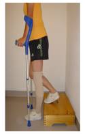
Beim Hinaufsteigen der Treppe geht das nicht-betroffene Bein voraus (Bild A).
 B
B
Beim Hinuntersteigen der Treppe geht das betroffene Bein voraus (Bild B)
Um möglichst schnell ein ergonomisches Gangbild zu erreichen, werden wir mehrmals Ihr Gangbild analysieren, um persönliche Korrekturen anzubringen und dazu angepasste Übungen instruieren.
Bis 3 Wochen nach der Operation ist unser Ziel, die Schwellung des Knies zu lindern, die passive Beweglichkeit zu steigern und die Stabilität des Knies zu verbessern, indem die Oberschenkelmuskulatur aktiv eingesetzt wird.
In der 3. und 4. Woche nach der Operation ist das Ziel, die vollständige Abschwellung des Gelenkes, die aktive und passive Beweglichkeit in Beugung und Streckung verbessern, sowie die Entwöhnung der Gehstöcke und die Verbesserung des Gleichgewichtes.
In der 5. und 6. Woche trainieren sie vor allem die Kraftausdauer der gesamten Beinmuskulatur. Dazu kombinieren wir gleichzeitig die bis jetzt erreichte aktive Beweglichkeit des operierten Beines.
Ab der 7. Woche nach der Operation ist das Ziel, die Muskulatur zu kräftigen und eine optimale Stabilität und Koordination zu erlangen z.B. beim Springen und Hüpfen.
Nach der 12.-24. Woche erfolgt die Reintegration in den Alltag (Hobby, Beruf...). Ein wettkampfspezifisches Aufbautraining für Sportler ist ab diesem Zeitpunkt wieder möglich:
Nach 4 Mt:
Lauf-, Wasser Jogging- und Fahrradsport
Nach 6 Mt:
Langlauf (klassisch), Schwimmen
Nach 9 Mt:
Langlauf (Skating), Skifahren, Snowboard, Eishockey, Fussball, Tennis, Volley- und Basketball
2.4.2.4.1 Nachbehandlungsschema für die stationäre Physiotherapie nach VKB-Plastik (mit Semitendinosus/ Gracilis Double Loop Technik)
Zeit |
Hauptziele |
Physiotherapeutische Massnahmen |
Präops- |
|
|
0ps-Tag |
|
|
1.postops- |
passiv
|
(2x/ 1h frei, nach Massgabe der Beschwerden) |
2.postops- |
passiv/assistiv/aktiv
Mantelspannung
|
|
ab 3. |
passiv/ assistiv/ aktiv
|
|
*solange bis eine gute M.Quadricepsinnervation ausgeführt wird bzw. die Schwellung abgeklungen ist.
Allgemein: Hierbei handelt es sich um eine isolierte VKB-Plastik-Nachbehandlung.
Falls weitere intraoperative Massnahmen getroffen wurden, wird die Nach-behandlung vom Operateur angepasst werden.(Bsp. Meniskusnaht lat. rechts).
Akute VKB-Läsionen werden schnell und progressiv rehabilitiert, chronische VKB-Läsionen dagegen langsamer rehabilitiert. Da dieses Konzept nicht überall bekannt und dementspre-chend nicht überall umgesetzt wird, müssen sowohl der Patient als auch die nachbehandelnde Physiotherapie auf die progressive Rehabilitation nach Rekonstruktion einer akuten VKB-Läsion aufmerksam gemacht werden.
2.4.2.4.2 Nachbehandlungsschema für die ambulante Physiotherapie nach VKB-Plastik (mit Semitendinosus/Gracilis Double Loop Technik)
Wundheilung |
Zeit |
Hauptziele |
Physiotherapeutische Massnahmen |
Entzündungs- |
Postop. |
|
|
Konsolidierungs- |
Postop. |
|
Ergänzend:
(geschl. Kette)
|
Postop. |
|
Ergänzend:
|
|
Reorganisations- und Umbauphase: |
Postop. |
|
Ergänzend:
Sprungaufbau:Squat-/ Countermouvement-/ Dropjump |
|
Postop. |
|
|
*solange bis eine gute Quadricepsinnervation ausgeführt wird bzw. die Schwellung abgeklungen ist. |
|||
